The fastest detecting and most advanced electron microscope in the world is coming to UCF’s Department of Physics, thanks to researchers at the university.
The group researches properties of materials that could be used in a variety of fields from medicine to engineering. There is the potential of discovering novel ways to deliver medicine in non-intrusive ways and creating better and faster semiconductors to help with communications and energy use. But to get there, researchers have to be able to observe the superfast processes happening in the material, which happens at subatomic levels.
The new microscope, the first to be located in the Western Hemisphere, will help researchers study the ultra-fast dynamics of electrons inside the materials.
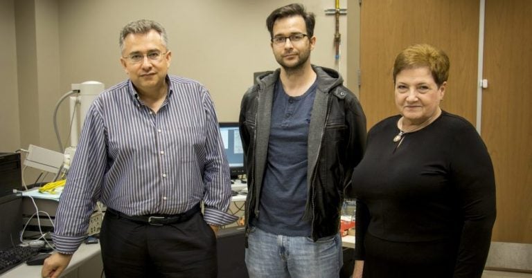
The team, led by physics Professor Leonid Chernyak, succeeded in landing a $1.5 million National Science Foundation grant to purchase the Allalin 4027 Chronos Cathodoluminescence Electron Microscope.
“This microscope can study a wide range of materials, from biomaterials to metals,” Chernyak said. “We predict it will help us see some new things that we hope lead to some big discoveries.”
When Chernyak came to UCF in 1999, his lab housed the first electron microscope in the department. The university now maintains several of them, but none with the sophistication of the Attolight (https://www.attolight.com) microscope. A team will install the new instrument next spring.
The current microscopes produce images by using electrons. A machine sends electrons down through its column to hit the sample being observed. When the electrons hit the sample, some so-called secondary electrons are created and detected, which produce pixels. These pixels create an image to be viewed and studied. These pixels create an image to be viewed.
The new electron microscope works similarly. However it is more sophisticated in that its detectors can be tuned for signals other than electrons – such as light.
The microscope can produce light by hitting the material with an electron beam, and the stimulated material then produces light. This process is called cathodoluminescence.
The microscope’s temperature can be varied over a wide range, allowing researchers to probe samples under a variety of conditions, explained the researchers.
The new machine can produce not just a continuous beam, but a pulse, which is not something most microscopes can do.
Chernyak explained that if the processes in the material are fast, then the beam shined on it has to be faster or you won’t see a reaction. The new machine is able to produce a pulse of 30 picoseconds. A picosecond is one trillionth of a second. The material’s response is then produced in nanoseconds, one billionth of a second.
“The ability to pulse is what sets this new machine apart,” said Elena Flitsiyan, associate lecturer and undergraduate program director in the department. “To technically make this happen is very challenging.”
Flitsiyan, who received her doctorate in physics and math from Moscow State University, said the pulsing beam can only be created by Attolight, the manufacturing company.
“The camera that detects its reaction can be described as a movie camera for the nano-world,” she said. “The microscope generates high-resolution images of samples and the ultrafast optics allow us to collect emitted light with high resolution.”
There are many applications, Flitsiyan added. The tool will give the scientists a way to see how currents propagate in a sample. Then they will be able to determine how imperfections affect electric charge carriers in semiconductors. These are essential materials for building electronic devices.
From space to medicine, this microscope will allow researchers across the university and the nation to discover new components in the materials they are studying.
Chernyak’s research is focused on semiconductors and Flitsiyan’s focus is on nuclear and radiation physics.
Additional investigators on the project include: Pieter Kik, Ph.D., and Romain Gaume, Ph.D., associate professors in the College of Optics & Photonics, who work with optical photonics and filters; Andre Gesquiere, Ph.D., an associate professor in the Department of Chemistry and the Nanoscience Technology Center, who works with biological materials; and Dmitry Kolpashchikov, Ph.D., an associate professor in the Department of Chemistry, who researches cancer cells.
Physics graduate student Jonathan Lee will be the primary user of the microscope as he studies semiconductors for his research.
In addition to aiding these researchers, the microscope will be available to other UCF researchers and students and collaborators at other universities and organizations in North and South America. External users will need to pay a fee to book time on the machine. The money collected will be used to service and maintain the instrument, Chernyak said.
“We expect great things,” Chernyak said. “We are counting the days until installation.”